
Imaging-Guided Precision PCI with OCT & IVUS in Mohali
Advanced Heart Care with Unmatched Precision
When it comes to treating complex coronary artery disease, precision matters. At her advanced cardiology practice in Sector 63, Mohali, Dr. Honey Sharma, a leading Interventional Cardiologist, offers Imaging-Guided Percutaneous Coronary Intervention (PCI) using the latest OCT (Optical Coherence Tomography) and IVUS (Intravascular Ultrasound) technologies.
These cutting-edge tools provide detailed, real-time imaging inside the arteries, enabling accurate diagnosis, perfect stent placement, and improved long-term outcomes for patients with coronary artery blockages.
What is Imaging-Guided PCI?
Percutaneous Coronary Intervention (PCI), commonly known as angioplasty with stent placement, is a minimally invasive procedure to open narrowed or blocked coronary arteries.
Imaging-guided PCI takes this a step further by using advanced imaging modalities such as:
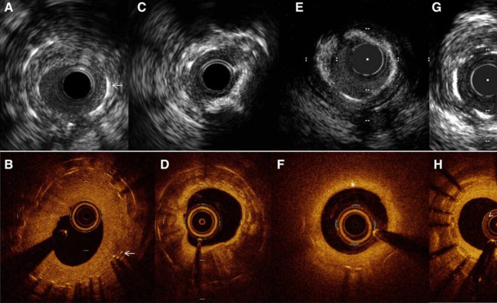
OCT (Optical Coherence Tomography) – Uses light waves to capture high-resolution images inside the artery.
IVUS (Intravascular Ultrasound) – Uses sound waves to provide a detailed cross-sectional view of the artery.
These technologies give Dr. Honey Sharma a clear and precise view of the artery’s interior, plaque characteristics, and stent positioning—something traditional angiography alone cannot achieve.
Why OCT & IVUS Matter in Heart Treatment
While standard angiography shows a silhouette of the coronary arteries, OCT and IVUS provide true-to-life, microscopic-level images. This means:
Exact measurement of artery size and blockage severity
Identification of plaque type (soft, calcified, or mixed)
Accurate stent sizing and placement
Prevention of stent under-expansion or malposition
Early detection of complications like dissection or thrombus
With this level of precision, procedural success rates are higher and the risk of repeat procedures is reduced.
Conditions Treated with Imaging-Guided PCI
Imaging-guided PCI is particularly beneficial for:
Complex coronary lesions (Left Main, Bifurcation, and Ostial lesions)
Heavily calcified blockages requiring Rota-Ablation or IVL
Chronic Total Occlusions (CTOs)
Small vessel disease
Patients with previous stents needing re-evaluation
Uncertain angiographic findings where exact plaque structure is unclear
Step-by-Step Process of Imaging-Guided PCI
1. Pre-Procedural Assessment
Detailed medical history
ECG and Echocardiogram
Coronary Angiography to identify potential blockages
2. Imaging with OCT or IVUS
A miniature imaging probe is inserted through the catheter into the coronary artery.
OCT captures high-resolution, light-based images.
IVUS captures ultrasound-based cross-sections of the vessel.
3. Guided Angioplasty & Stenting
Precise stent selection based on measurements from OCT/IVUS.
Balloon angioplasty to open the blockage.
Stent deployment with imaging confirmation for optimal expansion.
4. Post-Procedure Imaging
Final OCT/IVUS scan ensures perfect stent placement and absence of complications.
Benefits of Imaging-Guided PCI with Dr. Honey Sharma
1. Higher Precision & Safety
Every millimeter counts in heart procedures. Imaging guidance ensures accurate stent sizing, reducing the risk of future complications.
2. Better Long-Term Outcomes
Properly placed stents mean fewer repeat procedures and better artery healing.
3. Expertise in Complex Cases
Dr. Sharma’s vast experience in complex coronary interventions makes her the preferred choice for high-risk angioplasties in Mohali and the surrounding region.
4. Minimal Recovery Time
With transradial access (wrist route), patients recover faster, experience less discomfort, and can return to daily life quickly.
OCT vs IVUS – Which is Better?
Both OCT and IVUS are excellent imaging tools, but each has unique strengths:
| Feature | OCT | IVUS |
|---|---|---|
| Image clarity | Very high (microscopic) | High |
| Penetration depth | Moderate | Deep |
| Best for | Fine structural detail, stent expansion check | Plaque composition, vessel size assessment |
Dr. Honey Sharma chooses the right modality based on the patient’s condition to achieve the best possible outcome.
FAQs on Imaging-Guided PCI in Mohali
1. Is OCT/IVUS imaging safe?
Yes, both are extremely safe and routinely used in advanced cardiac centers worldwide.
2. Does it increase the procedure time?
Only slightly, but the benefits in terms of precision far outweigh the extra few minutes.
3. Will it cost more than regular angioplasty?
There may be a slight cost difference, but it reduces the risk of repeat procedures—saving money and improving health in the long run.
4. Can all patients undergo imaging-guided PCI?
Most can, and it is especially recommended for complex or unclear cases.
5. Is it available everywhere in Mohali?
No—OCT and IVUS require specialized equipment and expertise. Dr. Honey Sharma is among the few cardiologists in the region offering this advanced service.
Why Mohali Patients Choose Dr. Honey Sharma for Imaging-Guided PCI
DrNB in Interventional Cardiology from Fortis Hospital, Mohali
8+ years of experience in coronary interventions
Expertise in Rota-Ablation, IVL, and complex lesion treatment
Member of CSI and WINCARS
Committed to holistic heart care
Book Your Appointment Today
If you have been diagnosed with a coronary blockage or are experiencing chest pain, shortness of breath, or fatigue, early treatment can save your life.